
The human male reproductive system consists of a group of sex organs that play a role in the process of human reproduction.
These organs are located outside of the body and within the abdomen (pelvic region).
The male reproductive system is responsible for sexual function, producing sperm, and semen, and helps in the transportation of semen and urine.
The male reproductive system is divided into 2 parts
- External genital organs – penis, scrotum, testicles
- Internal genital organs – vas deferens, sperm ducts, urethra, accessory ducts.
Anatomy of the male reproductive system
External genital organs
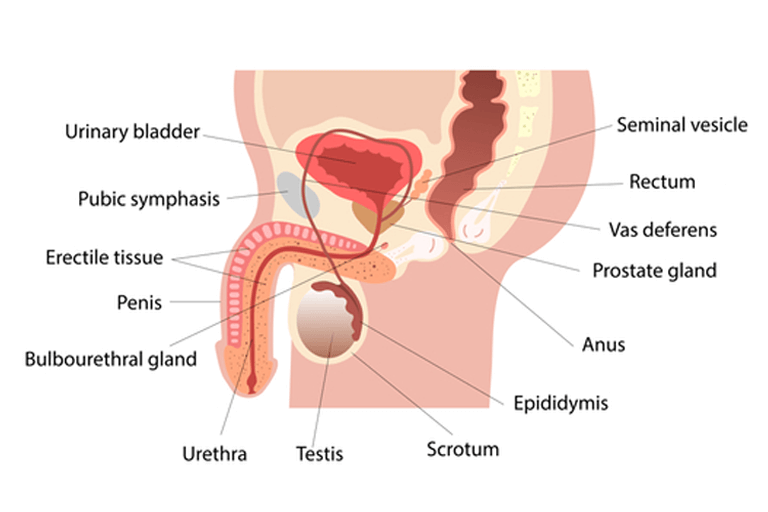
Penis
The penis is a male organ for sexual intercourse and urination. It has 3 parts
1. Root
- The root is the base of the penis and attaches to the abdomen’s wall.
2. Body (shaft)
- The shaft has a cylinder or tube-like shape
- It consists of 3 internal chambers. Two larger chambers (corpus cavernosa) run side by side over the shaft. The third chamber (corpus spongiosum) surrounds the urethra (urinary duct).
- There is a special sponge-like erectile tissue inside these three chambers.
- The erectile tissues contain thousands of spaces.
- During a sexual erection, the spaces fill with blood, penis becomes hard (rigid) and ready for sexual activity.
- When the penis is erect, larger chambers of the penis press against the part of the urethra where urine flows. This blocks urine flow so that only semen can ejaculate.
3. Glans (Head of penis)
- The glans is the cone-shaped (enlarged bulbous-shaped) tip of the penis.
- A loose and stretchy layer of skin (foreskin) covers the glans, which changes its size during erection.
- The average size of the peins is about 3.5 inches when it is soft and about 5 inches when erect.
Scrotum
– The scrotum is a loose pouch-like sac of the skin that hangs behind the penis.
– It holds and protects the testicles and provides a sort of “climate control system” for normal sperm development.
– It also contains numerous nerves and blood vessels.
– During times of lower temperature in the surrounding atmosphere, the cremaster muscle contracts and pulls the scrotum near the body while dartos muscles give it a wrinkled appearance; when there is an increase in surrounding temperature, cremaster muscles relax which brings down scrotum away from the body, and dartos muscles remove wrinkles. Thus by this, they maintain an appropriate temperature for sperm production.
– The testicles are one of two oval-shaped organs that lie in the scrotum and produce sperm and male sex hormones.
– Each testicle is about 5 cm long.
– Each testicle is covered by tough fibrous layers of tissue called tunica.
The outer layer is called tunica vaginalis.
The inner layer is called tunica albuginea.
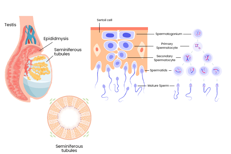
– The testicle is divided into lobules. Each lobule has tiny U-shaped tubes called seminiferous tubules tightly coiled within each testicle.
– The seminiferous tubules open into a series of uncoiled interconnected channels called rete testis. Rete testis are further connected to a tightly coiled tube (epididymis) which is in turn connected to vas deferens (a long large spermatic cord).
– Each testicle is held in the scrotum by a spermatic cord. Each spermatic cord is made up of tough connective tissues, muscle, vas deferens, arteries, veins, lymph vessels, and nerves.
– Testicles have two main types of cells (germ cells and stromal cells) that produce sperm and male sex hormones.
1. Germ cells
- Germ cells lie in seminiferous tubules. The process of making sperm starts in germ cells.
- Germ cells repeatedly divide and further mature into sperm.
- As germ cells mature into sperm cells, they move through seminiferous tubules and to the epididymis.
- The epididymis stores sperm so they can completely mature.
- Mature sperm cells travel through the vas deferens during ejaculation.
- Along the way, seminal vesicles and prostate gland secretions mix with sperm to create semen.
- The semen is pushed out of the body through the urethra during ejaculation. Sperm in the semen can fertilize a female egg (ovum) to start pregnancy.
2. Stromal cells
- The seminiferous tubules in the testicles contain several types of stromal cells, including Sertoli cells, Leydig cells, and myeloid cells.
– Sertoli cells: These highly branched cells line the seminiferous tubules and regulate spermatogenesis.
– Leydig cells: They produce testosterone hormone, which helps germ cells make sperm and the reproductive organs develop and function.
– Myeloid cells: These squamous contractile cells of the seminiferous tubules generate peristaltic waves.
Internal genital organs
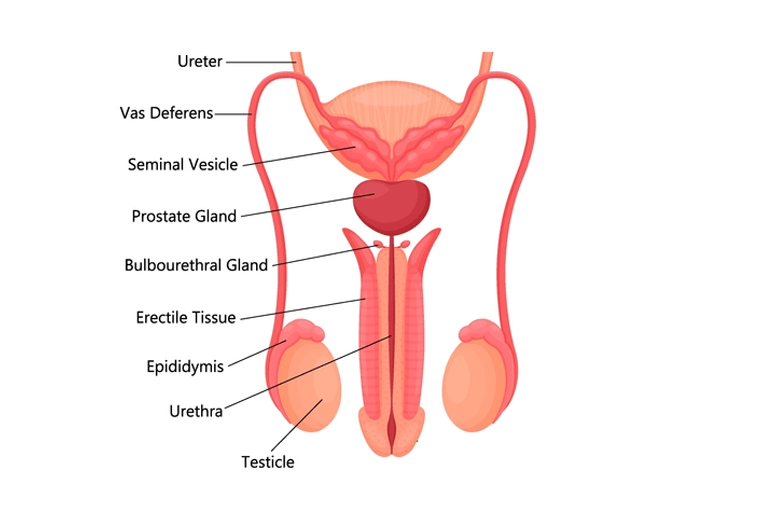
Vas deferens
The vas deferens is a long muscular tube that travels from the epididymis into the pelvic cavity, just behind the urinary bladder.
The vas deferens transports mature sperm to the urethra.
Urethra
The urethra is a tube that carries urine from the bladder and ejaculates semen when you reach orgasm.
Accessory glands
Seminal vesicles:
- The seminal vesicles are sac-like pouches that attach to vas deferens near the base of the bladder.
- They make up to 80% of seminal fluid.
Prostate gland:
- The prostate is a walnut-sized gland.
- The prostate adds additional fluid which helps nourish sperm.
Bulbourethral glands
- These glands are pea-sized on the sides of the urethra, just below the prostate.
- They create a clear, slippery fluid in the urethra.
- This fluid lubricates the urethra and neutralizes an acid that may remain in urine.